
The practice of dentistry has historically been defined by tactile skill, visual evaluation, and manual dexterity. For generations, practitioners relied on physical impressions, two-dimensional radiographs, and steady hand-eye coordination to diagnose oral pathologies and execute treatments.
While these foundational skills remain essential, the limits of human sight and physical materials inherently introduce minor variables into clinical outcomes.
Modern dentistry has undergone a massive paradigm shift, moving rapidly away from analog techniques toward an era of computational accuracy.
Today, advanced digital tools serve as an objective extension of the dentist’s hands and eyes. By digitizing the oral cavity, clinicians can plan complex interventions with microscopic precision, eliminate human error, and deliver highly predictable treatment outcomes that were technically impossible just a few decades ago.
Revolutionizing Diagnostics with High-Definition Spatial Mapping
Every successful dental intervention depends on a flawless diagnosis. Traditional imaging methods frequently force clinicians to make treatment assumptions based on flattened, shadowed perspectives of three-dimensional structures.
The Shift to Three-Dimensional Cone Beam Volumetric Imaging
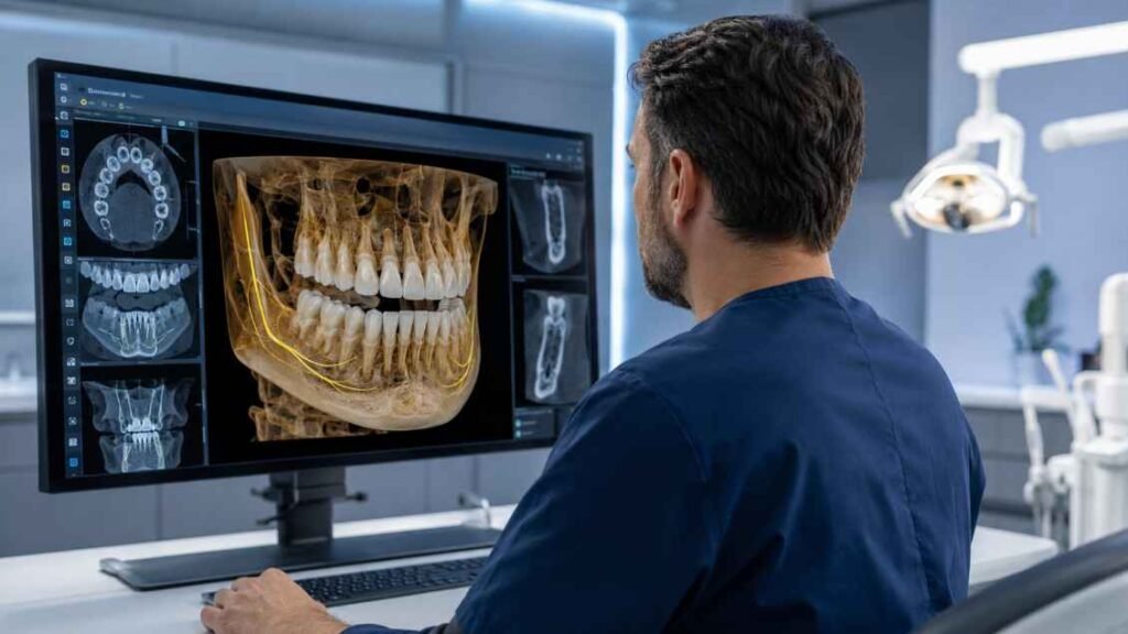
Standard two-dimensional periapical and panoramic X-rays are highly prone to structural overlap, magnification errors, and distortion. Cone Beam Computed Tomography has revolutionized diagnostic capabilities by capturing thousands of data points in a single, rapid scan.
This technology constructs a highly accurate, three-dimensional digital model of the patient’s craniofacial anatomy, including bone density, tooth root orientations, and nerve pathways.
Having access to this depth of anatomical data allows dentists to evaluate implant sites, identify hidden root fractures, and detect periapical lesions long before they manifest on traditional films. This early detection capability prevents exploratory surgeries and allows for highly conservative tissue management.
Eliminating the Inaccuracies of Analogue Dental Impressions
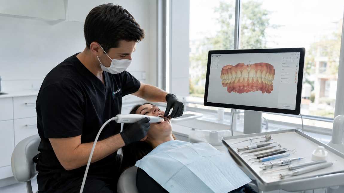
Intraoral scanners have systematically replaced the messy, uncomfortable alginate and polyvinyl siloxane molds of the past. These wand-like digital cameras capture thousands of surface images per second, assembling a perfect virtual replica of the teeth and gingiva in real time.
Many patients also consider how the products and materials used in their care affect overall health and the environment, which is why Eco-Friendly Skincare is relevant.
Analog materials are highly susceptible to tearing, warping during transit, and contracting as they set. Digital scans eliminate these physical vulnerabilities entirely, providing dental laboratories with micron-level surface data.
For persistent or unusual oral lesions, Mouth Sores When to Worry and When to Wait to understand when professional evaluation is necessary.
This absolute precision ensures that crowns, bridges, and veneers fit the prepared tooth structure perfectly, significantly reducing the need for chairside adjustments during the final cementation appointment.
Computer-Guided Restorative and Implant Procedures
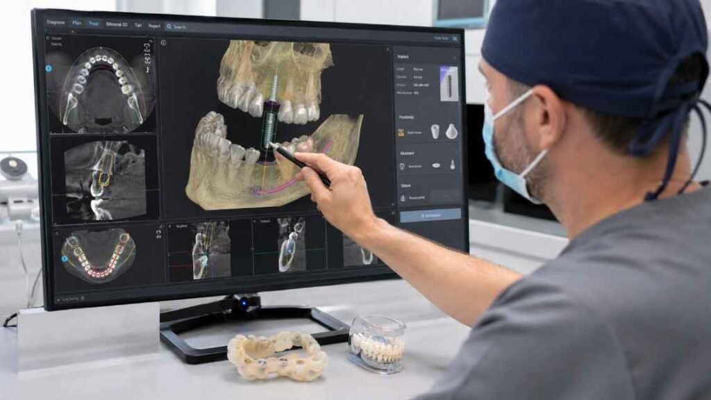
Once a digital map of the oral cavity is established, practitioners can use specialized software to simulate treatments virtually long before touching a physical handpiece.
Prosthetically Driven Implant Placement
In the past, dental implants were often placed freehand, relying strictly on the surgeon’s visual assessment of the open bone site. Today, modern surgical planning software allows dentists to merge intraoral surface scans with bone data.
Practitioners can virtually position an implant on the computer screen, adjusting the depth, angle, and rotation to maximize bone support while completely avoiding vital structures like the inferior alveolar nerve or maxillary sinus.
Once the virtual design is finalized, a custom surgical guide is manufactured via 3D printing. Pinning this guide securely over the patient’s teeth during surgery restricts the drill path completely, forcing the implant into the exact pre-planned position with millimeter precision.
Advanced Automation in Orthodontic Case Setups
The pursuit of clinical accuracy has also significantly transformed the field of orthodontics, particularly in the placement of traditional and aesthetic bracket systems.
Historically, bonding brackets directly to teeth chairside was a labor-intensive process where minor visual miscalculations could lead to improper root torque or unwanted tooth movement, necessitating extensive wire bending later in treatment.
Modern workflows bypass this challenge by utilizing sophisticated digital indirect bonding software to virtually position brackets on a high-definition 3D model of the patient’s dental arches.
The computational system simulates tooth movement and models wire progressions, allowing the clinician to fine-tune the bracket heights and angles without any chairside pressure.
Once approved, the software designs a custom, form-fitting transfer tray that is 3D printed in-house or at a specialized lab. This tray holds the physical brackets in their exact digital configurations, allowing the clinical team to bond an entire arch of braces flawlessly in a fraction of the traditional chairside time.
Artificial Intelligence and Real-Time Secondary Assessments
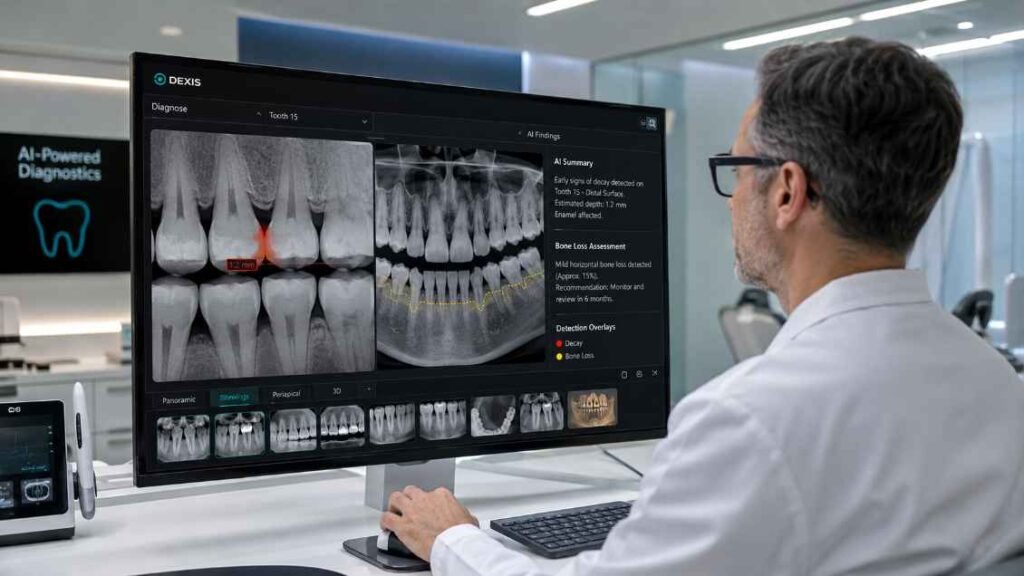
The integration of artificial intelligence into dental workflows serves as an invaluable diagnostic safety net, mitigating the risks of fatigue and human oversight during busy clinical schedules.
Objective Radiographic Analysis
AI-powered diagnostic software acts as an automated, highly objective second opinion. As soon as a digital radiograph is uploaded, the cloud-based algorithms scan the image instantly, comparing the grayscale densities against databases containing millions of validated dental conditions.
The software automatically highlights early-stage interproximal decay, marginal discrepancies around existing restorations, and subtle bone loss using bright, color-coded visual markers. This validation system ensures that emerging pathologies are caught early, protecting patients from advanced infections and proving that modern tech can elevate standard diagnostic accuracy.
By fully embracing the digital ecosystem, modern dental practices are successfully replacing subjective human estimations with verifiable, repeatable digital precision, raising the standard of oral healthcare to unprecedented heights.